Category: People
Nancy Berliner, MD
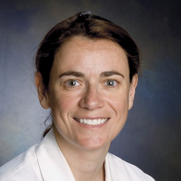
Alexandra J. Golby, MD

Alexandra Golby’s research focuses on the translation of a broad range of neuroimaging techniques to neurosurgical planning and intraoperative guidance. The overarching goal of this work is to help surgeons perform optimal brain surgery by defining and visualizing critical brain structures and pathologic tissue to be removed.
She was the recipient of a Brigham Research Institute Shark Tank Award in the amount of $50,000 for her project with Pablo Valdes Quevado, MD, PhD, ‘Color Coding Tumors’ in 2018.
Since 1998, she has worked on the development and validation of fMRI for the pre-operative evaluation of patients with lesions in and near critical areas of the brain. This has been a translational research effort which adapted fMRI, initially developed as a neuroscience technique to be applied in groups of subjects to make statistical inferences about populations, to the vastly different scenario of clinical decision-making for individual patients. Since the research program began at the Brigham in 2003, she has developed new techniques for the use of fMRI in single subject analyses necessary for surgical planning. In addition, she has developed numerous acquisition strategies geared towards accommodating the limited neurologic functions of some patients as well as analytic approaches to maximize the utility of fMRI for surgical planning.
Presurgical fMRI has the potential to bring meaningful pre-operative individualized functional anatomy mapping to neurosurgeons around the world as an alternative to awake mapping, a technique which is demanding and remains limited to very specialized centers.
Alexandra J. Golby, MD, Department of Neurosurgery
I have also worked extensively on the translation of diffusion MRI (dMRI) including tensor imaging (DTI) to map white matter anatomy in neurosurgical patients. Diffusion MRI allows the in vivo depiction of the location, course and integrity of macroscopic white matter tracts in the brain through a process known as tractography. As with fMRI, the translation of this technology to clinical decision-making has required numerous fundamentally novel approaches. We have developed segmentation approaches for defining tracts based on high dimensional clustering as well as statistical atlases which allow labeling of individual patient tracts even in the setting of mass effect and peritumoral edema.
Her group works collaboratively with MRI physics and MRI analysis groups to continue to be at the forefront of technical innovation. They have released many of our tools to the public via 3D Slicer (slicer.org) and we have organized several international challenge workshops to apply diffusion techniques to real world clinical data.
With both these methods, translation of the technology required understanding of clinical needs, constraints, and opportunities for improved clinical care. Specific analysis techniques needed to be developed to adopt these techniques so that they were applicable to single subject data, and, in particular, to neurologic patients who have structural lesions and often are limited by their neurological deficits. In these efforts, her group works closely and collaboratively with scientists in radiology and computer science to translate emerging technical innovations into the operating room.
Another major area of translational investigation is in the development of intraoperative imaging techniques. Alexandra was the lead surgeon in developing the AMIGO (Advanced Multi-modality Image-Guided Operating Suite) at the Brigham and serve as the Co-director of AMIGO. AMIGO is one of the key resources of the National Center for Image Guided Therapy funded by NIH. This suite contains all contemporary imaging methods within an operating room environment and was specifically designed to support translational research. The suite is the site of many of surgical procedures in which they are developing strategies for intraoperative imaging and guidance.
Several of her group’s important research efforts are built on the AMIGO platform. These include the intra-operative use of high field MRI including development of intra-operative dMRI tractography. We have also leveraged the resources of the AMIGO suite to develop novel strategies to simplify intraoperative imaging using techniques such as ultrasound and stereovision to give surgeons information in near real time to guide surgery. Another area of research leveraging the resources of AMIGO is the development of tissue level molecular imaging. They have funded collaborative projects using mass spectrometry, Raman spectroscopy, and fluorescence imaging.
Our eventual goal is to give surgeons in most settings tools that will help them to perform safer and more effective surgery.
Alexandra J. Golby, MD

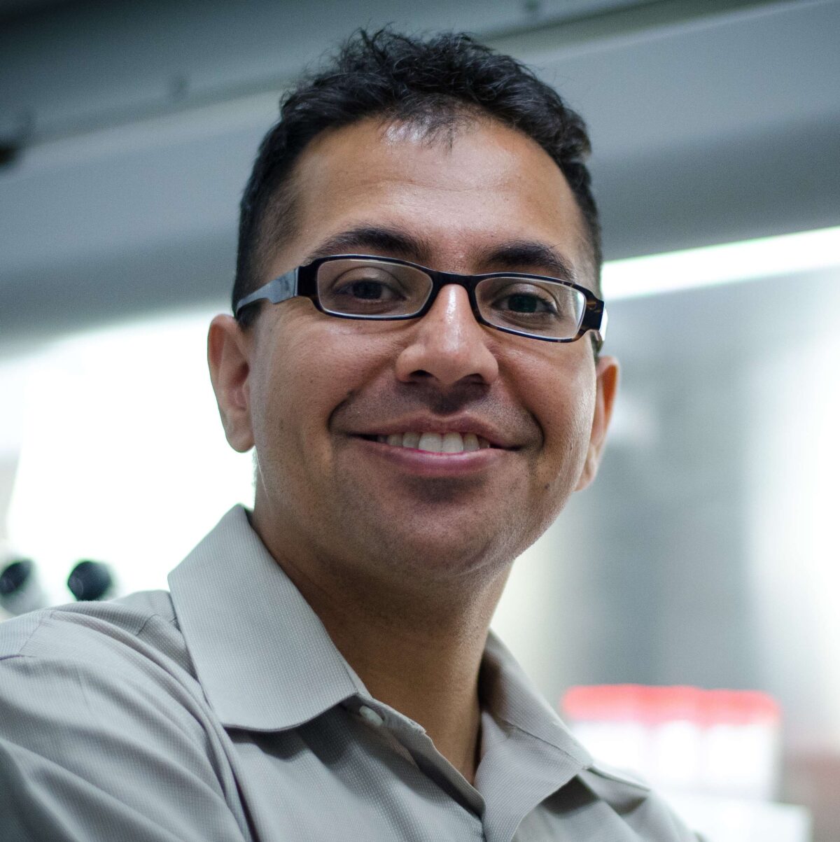
Vikram (Vik) Khurana is on the Neurology faculty at Harvard Medical School. He is currently the Chief of the Movement Disorders Division at Brigham and Women’s Hospital and a principal research investigator in the Ann Romney Center for Neurologic Diseases. He is also Principal faculty at the Harvard Stem Cell Institute, an Associate Member at the Broad Institute of Harvard and MIT, and a Robertson Stem Cell Investigator of the New York Stem Cell Foundation. In 2014, Vik co-founded the biotech company Yumanity Therapeutics, where he currently serves as Senior Advisor.
Vik’s clinical and research interests relate to neurodegenerative movement disorders, including Parkinson’s disease, multiple system atrophy, progressive supranuclear palsy and the cerebellar ataxias. He grew up in Sydney, Australia, and is a medical graduate of the University of Sydney. He came to Boston as a Fulbright Scholar in 2001, obtaining his Ph.D. in neurobiology from Harvard University in 2006. His dissertation adviser was Dr Mel Feany in the Department of Pathology at Brigham and Women’s Hospital. He completed his residency in neurology at Brigham and Women’s and Massachusetts General Hospitals, and Fellowship training in movement disorders and ataxia at Massachusetts General Hospital with Drs Jeremy Schmahmann, John Growdon and Lew Sudarsky. During that time, he was awarded grants from the American Brain Foundation, Parkinson’s Disease Foundation, Multiple System Atrophy Coalition and Harvard Neurodiscovery Center.
Vik received postdoctoral scientific training in the laboratories of Drs Susan Lindquist and Rudolf Jaenisch at the Whitehead Institute, where, along with his wife Chee Yeun Chung, he led a study that succeeded in identifying and reversing pathologies in stem cell models of Parkinson’s disease (Chung*, Khurana* et al. Science 2013). As you will see on this website, Vik’s current research efforts continue toward development of therapies for neurodegenerative diseases by using models as simple as Baker’s yeast cells, to much more complex human stem cell- and organoid-based models. He is thrilled to have such a great team to join him on this quest.
Once upon a time Vik enjoyed playing blues guitar, photography and long-distance kayaking. These days, in his spare time he struggles to keep up with his two young daughters. Vik would be quite incapacitated without the constant support of his life and scientific partner, Chee Yeun, and his extended family in Australia.